
Projects
Various projects from the archive
-

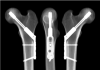
Virtual x-rays of the 3-hole Austofix Tectona sliding hip screw. Implant is shown with the middle screw removed in two independent planes (coronal and sagittal)
-
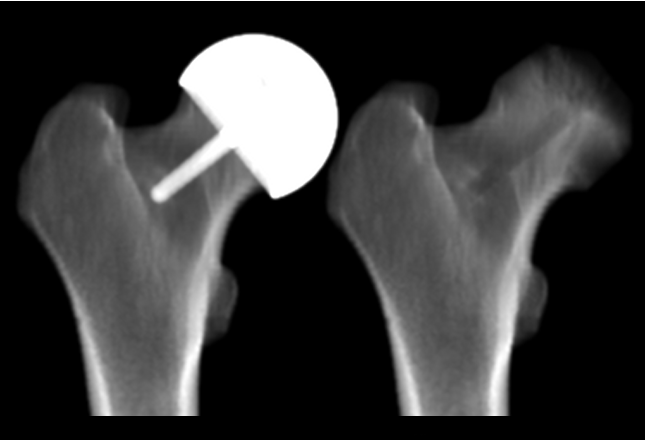
Virtual x-rays of the Birmingham Head Replacement (BHR) hip resurfacing implant with implant (left) and without implant (right). The latter can be used to determine the change in bone density beneath the head, normally obscureed by the implant.
-
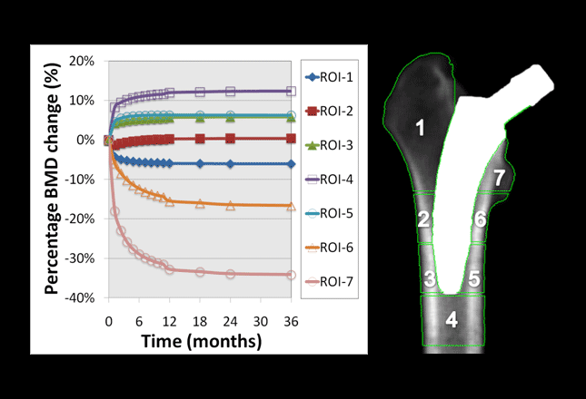
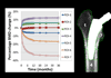
Example of how virtual x-rays are used to analyse results from a bone remodelling analysis. Implant shown is the MiniHip conservative hip stem (Corin Group PLC). Regions of Interest (ROIs) 6 and 7 display the greatest degree of bone resorption
Virtual x-rays and bone remodelling
Background: The implantation of a medical implant, such as a hip or knee replacement, into bone leads to a change in mechanical loading and invokes a bone remodelling response in an attempt to bring the system back into equilibrium. Any excessive reduction in bone density may lead to loosening and failure of the implant.
Clinical studies typically use Dual Enery X-Ray Absorptiometry (DEXA) to evaluate changes in Bone Mineral Density (BMD). In such studies, DEXA images are taken pre-operatively and then post-operatively approx every 6-12 months. These images are then compared and the changes in BMD over time for a number of Regions of Interest are established. Although clinical studies using DEXA play an important role in the evaluation of the long-term performance of medical implants, they cannot be used to predict the long-term outcome of a device.
Virtual x-rays: Numerical bone remodelling analyses together with “virtual x-rays” have been used successfully to predict the long-term clinical outcome of many implants while still in the design phase. The bone remodelling algorithms used by MDR have been validated using clinical DEXA results, made possible by the “virtual x-rays” outputted as part of each analysis. This sets MDR apart from many other researchers that do not output “virtual x-rays” and are therefore unable to validate their numerical models using clinical data.
The “virtual x-rays” are created by “mapping” the bone densities from the numerical model onto the three planes of a specified coordinate system, such that x-rays in any desired plane are able to be generated. Images can be created with or without the implant, such that areas normally obscured in DEXA images are visible in “virtual x-rays”.